Angiography is an essential medical imaging technique we use to visualize blood vessels. By injecting a radio-opaque dye into the bloodstream, we can create detailed images that help us diagnose cardiovascular conditions. This non-invasive procedure markedly enhances our ability to assess blood vessel health and identify issues like blockages early on. Understanding the nuances of angiography can provide valuable insights into its significance and application in modern medicine, revealing much more than meets the eye.
Main Points
- Angiography is a medical imaging technique used to visualize blood vessels and organs by injecting a radio-opaque dye into the bloodstream.
- It aids in diagnosing vascular disorders and detecting blockages, critical for preventing strokes and heart attacks.
- The procedure involves catheter insertion and contrast dye injection, allowing detailed X-ray imaging for accurate assessments.
- History of angiography includes significant advancements, such as Egas Moniz’s cerebral angiography in 1927 and selective coronary angiography discovered by Mason Sones in 1958.
- Various imaging techniques, including X-ray, CT, and MRI angiography, enhance diagnostic capabilities and patient outcomes through improved visualization.
Definition of Angiography

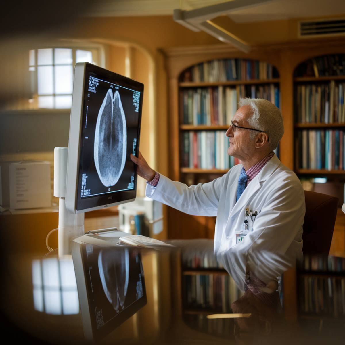
When we think about understanding our body’s vascular system, angiography stands out as a pivotal medical imaging technique. This specialized process allows us to visualize the inside of blood vessels and organs using a contrast agent and X-ray imaging. By injecting a radio-opaque dye into the bloodstream, we can create detailed images—known as angiograms—of how blood flows through arteries, veins, and heart chambers.
The benefits of angiography are significant; it aids in diagnosing disorders affecting blood vessels, detecting blockages, and guiding treatment decisions. Moreover, the introduction of the Seldinger technique in 1953 has significantly enhanced safety during catheter insertion. Additionally, this procedure is often performed as an outpatient procedure, ensuring patient comfort and convenience. To further emphasize its importance, angiography assesses blood vessel health and plays a crucial role in identifying potential risks such as stroke or heart attack. However, we must also remain mindful of the challenges. The procedure can carry risks such as allergic reactions to the contrast dye or complications from catheter insertion. Despite these challenges, angiography remains an invaluable tool in modern medicine, enhancing our ability to understand and manage vascular health effectively.
History of Angiography
As we explore the history of angiography, we find ourselves tracing a remarkable journey that began in the early 20th century with Egas Moniz, who forged the path for cerebral angiography in 1927. His innovative technique of injecting a radiopaque substance into the bloodstream to capture X-ray images of blood vessels laid the groundwork for future angiography innovations. The contributions of the university and medical community in Lisbon further propelled this field forward.
Significant historical milestones include Reynaldo dos Santos’ first aortogram in 1929 and Werner Forssmann’s daring self-catheterization the same year. Fast forward to 1958, when Mason Sones accidentally discovered selective coronary angiography, revolutionizing cardiac diagnostics. The technique involved selective contrast dye injection into coronary arteries, allowing for accurate visualization of arterial blockages and abnormalities. The introduction of the Seldinger technique in 1953 made procedures safer, paving the way for the sophisticated Quantitative Coronary Angiography in the 1970s. Each stride marked a defining moment in our understanding of vascular health, making angiography an indispensable tool in modern medicine. Notably, angiography has since evolved to greatly assist in diagnosing diseases of blood vessels, emphasizing its role in identifying issues within the vascular system during procedures.
Importance of Angiography

Angiography stands as a cornerstone in modern medicine, vastly improving diagnostic accuracy for a range of vascular conditions. By providing essential insights into treatment planning and facilitating the early detection of diseases, it empowers both physicians and patients alike. As we explore its significance, we’ll uncover how this technique enhances our ability to address cardiovascular health with precision and foresight. Moreover, angiography reveals blood circulation through a specialized X-ray exam, offering crucial information that other imaging techniques may not provide. This non-invasive procedure allows for detailed imaging of blood vessels, which is especially valuable in diagnosing blockages and abnormalities in the coronary arteries.
Diagnostic Accuracy Improvement
In order to enhance our understanding of cardiovascular health, we must acknowledge the critical role angiography plays in improving diagnostic accuracy. By integrating advanced techniques and technologies, we’ve made significant strides in diagnostic error reduction and imaging accuracy enhancement. Key factors include:
- Enhanced contrast agents for clearer visualization
- Integration of AI-QCT in coronary CT angiography
- Real-time imaging capabilities for dynamic assessment
These advancements allow us to accurately diagnose conditions like significant stenoses such as atherosclerosis and embolisms, ultimately leading to better patient outcomes. Angiography is a vital procedure that helps identify blockages or narrow spots in blood vessels, resulting in more informed treatment decisions. We’re confident that continuous improvements in modalities and techniques will further refine our diagnostic practices, ensuring that we catch conditions earlier and with greater precision, paving the way for effective intervention.
Treatment Planning Assistance
Harnessing the diagnostic insights gained from enhanced angiography practices, we can effectively leverage these capabilities in treatment planning. The angiography benefits extend beyond mere visualization; they guide our decisions in managing conditions such as blocked or narrowed arteries. With procedures like angioplasty and stent placement directed by angiograms, we’re able to enhance treatment effectiveness considerably. Furthermore, by identifying abnormal blood vessels and planning interventions like embolization, we can tailor our approach to each patient’s unique needs. This process not only guarantees that we’re tackling the problem at its core but also improves the overall success of further procedures like coronary artery bypass grafting. Ultimately, angiography serves as a pivotal tool in achieving excellent patient outcomes.
Early Disease Detection
While we often take our vascular health for granted, the early detection of diseases like peripheral artery disease (PAD) is essential for preventing serious complications. Angiography plays a pivotal role in early screening, allowing us to identify blockages and hidden abnormalities before they escalate.
By employing angiography, we can benefit from:
- Timely interventions that mitigate severe outcomes like limb amputation
- Detailed images that help plan appropriate treatments for complex cases
- Identification of asymptomatic conditions, ensuring proactive disease prevention
This method not only visualizes our blood vessels but also underscores the importance of monitoring our vascular health. Early detection truly empowers us to safeguard against more severe vascular diseases.
How Angiography Works

When we think about how angiography works, it all begins with the careful insertion of a catheter into a targeted artery, often using local anesthesia for comfort. Once positioned, we inject a contrast agent that illuminates the blood vessels, creating a vivid roadmap for imaging. The detailed X-ray analysis that follows reveals critical insights, allowing us to understand vascular health with tremendous clarity.
Catheter Insertion Process
To successfully navigate the catheter insertion process during angiography, we must meticulously prepare both the patient and the site of insertion. This preparation includes several essential steps:
- Shaving and cleaning the insertion site with antiseptic fluid
- Administering local anesthesia for comfort
- Setting up ECG monitoring to track heart activity
Once the patient is comfortably positioned, we make a small incision in the groin or arm to access the artery. A sheath, or introducer, is inserted to facilitate the insertion of various catheter types. Using x-ray guidance, we carefully thread the flexible catheter through blood vessels, ensuring precise catheter navigation and minimal discomfort. Throughout the process, we continuously monitor critical signs, keeping the patient’s safety and comfort as our top priorities.
Contrast Agent Injection
In angiography, the role of contrast agent injection is essential, fundamentally enhancing our ability to visualize and assess blood vessels. We typically use iodine-based contrast for X-ray and CT scans, while gadolinium serves MRI purposes. The contrast agent is injected into veins or arteries through catheters, guaranteeing ideal distribution throughout the body. Employing automatic pumps allows us to control injection rates, which is vital for patient safety. We monitor for allergic reactions and potential complications, like compartment syndrome, continuously during and after the procedure. Careful pre-procedure preparation helps mitigate risks, such as eliminating certain medications. Ultimately, this meticulous process guarantees we gain the clearest insights into vascular health while prioritizing injection safety.
Imaging and Analysis
Following the injection of the contrast agent, we harness advanced imaging techniques to visualize and analyze blood vessels effectively. Using X-ray technology, we capture detailed images that highlight essential vascular structures, aiding in our angiography evaluation.
Key aspects of our imaging analysis include:
- Real-time guidance: We navigate the catheter with precision through the vascular system, guaranteeing ideal positioning.
- Distinct image characteristics: Different tissues’ absorption rates reveal variations in the images, allowing us to pinpoint abnormalities.
- Comprehensive assessment: The resulting angiograms enable us to detect issues like blockages or narrowing, driving informed treatment decisions.
Through these meticulous processes, we guarantee that the angiography evaluation provides significant insights for patient care.
Types of Angiography

While exploring the various types of angiography, we find that each method is tailored to address specific vascular concerns throughout the body. Conventional angiography shines in its functional angiography applications, enabling clear visualization of blood vessels after contrast dye administration. CT angiography and MRA have emerged as invaluable angiography technological advancements, offering non-invasive alternatives that yield detailed images with less patient risk.
Peripheral angiography focuses on arteries in the limbs, diagnosing conditions like peripheral artery disease. Cerebral angiography scrutinizes brain blood vessels, essential for detecting aneurysms and arteriovenous malformations. Meanwhile, pulmonary angiography effectively visualizes pulmonary vessels to uncover embolisms. Other methods like aortography assess the aorta, and fluorescein angiography evaluates retinal health. Each technique contributes to a thorough understanding of vascular health, allowing personalized treatments and interventions for various disorders.
Coronary Angiography
As we explore coronary angiography, we see it as an essential procedure that allows us to assess blood flow in the heart’s arteries using a specialized dye and X-ray technology. This diagnostic tool helps identify narrowed or blocked coronary arteries, enabling us to diagnose conditions like coronary artery disease effectively.
Key insights about coronary angiography include:
- Patient education is fundamental as it prepares individuals for what to expect during the procedure.
- Understanding coronary symptoms can lead to timely intervention, highlighting the necessity of seeking medical advice.
- Post-procedure care is critical for a smooth recovery, including monitoring the catheter insertion site.
During the procedure, we’re awake and feel minimal discomfort due to local anesthesia, and it usually lasts about 30 to 60 minutes. By applying this technology, we can plan further treatment options, promoting better heart health and patient outcomes.
Cerebral Angiography

Cerebral angiography stands as an essential tool in modern medicine, allowing us to visualize the intricate network of blood vessels in the brain with remarkable precision. Employing a minimally invasive approach, this procedure uses a contrast material combined with x-ray imaging to disclose cerebral conditions such as aneurysms and arteriovenous malformations. As we insert a catheter—often in the groin or wrist—we gain the ability to diagnose and treat in one seamless process, illustrating the benefits of contemporary imaging innovations.
Not only does cerebral angiography help confirm vascular issues and evaluate blood supply to tumors, but it also prepares us for surgical interventions by examining abnormalities. With careful preparation to guarantee safety, we can provide detailed images that inform critical decisions. While risks such as allergic reactions exist, the advances in endovascular treatment notably enhance our therapeutic options, making cerebral angiography a cornerstone in addressing complex neurological challenges.
Pulmonary Angiography
Pulmonary angiography serves as an important procedure for examining the blood vessels of the lungs, providing us with vital insights into various pulmonary conditions. This technique is particularly valuable for pulmonary embolism diagnosis, especially when other methods yield inconclusive results. Using fluoroscopy, we identify vascular abnormalities effectively.
Key applications include:
- Confirming pulmonary embolism and chronic thromboembolic pulmonary hypertension (CTEPH)
- Planning interventions like balloon pulmonary angioplasty
- Detecting vascular issues such as aneurysms or arteriovenous malformations
During the procedure, specialists—often interventional radiologists—insert a catheter and inject contrast dye to visualize lung blood flow accurately. We closely monitor the patient throughout, ensuring safety while gaining detailed information necessary for diagnosis and treatment planning. Ultimately, pulmonary angiography stands as an essential tool in delivering thorough care for pulmonary vascular concerns.
Renal Angiography
Understanding the intricate blood supply to our organs is essential for effective diagnosis and treatment, and renal angiography plays a key role in examining the blood vessels within the kidneys. This imaging test helps us identify conditions such as aneurysms, stenosis, and thrombosis that can affect renal health. During the procedure, we guide a catheter through a blood vessel to inject a contrast dye, allowing us to capture detailed X-ray images of renal blood flow.
Before we start, it’s important to discuss any allergies or medical conditions to guarantee procedural safety. We recommend staying hydrated after the procedure to flush out the dye. While complications are rare, awareness of risks like bruising or potential allergic reactions is significant. Renal angiography not only aids in diagnosing renal diseases but also refines our understanding of kidney function and treatment strategies, ultimately contributing to better patient outcomes.
Peripheral Angiography
As we explore peripheral angiography, it becomes evident that this specialized imaging technique is essential for diagnosing and treating conditions affecting the arteries in the extremities. By employing this approach, we can identify issues like peripheral artery disease (PAD), which commonly manifests through peripheral symptoms such as pain in the legs, cold skin, and unhealing sores.
Peripheral angiography is vital for diagnosing and treating extremity artery conditions, helping to identify issues like peripheral artery disease.
Here are some key aspects of peripheral angiography:
- Blockage Detection: It pinpoints arterial blockages in legs and feet.
- Risk Assessment: Individuals with high blood pressure, high cholesterol, diabetes, or a smoking history are particularly at risk.
- Treatment Options: Angioplasty and stent placement can occur alongside the imaging procedure.
Through these insights, we gain a better understanding of how peripheral angiography plays an essential role in diagnosing vascular conditions and facilitating timely interventions, ensuring better health outcomes for our patients.
The Angiography Procedure

While we prepare for the angiography procedure, it’s essential to recognize the careful orchestration involved in this diagnostic technique. We lie on a table in a specialized environment, often resembling an operating theater, with straps ensuring our safety. A small incision is made over an accessible artery, allowing a catheter to be inserted. As contrast dye is injected, it enhances blood vessel visibility on X-rays, facilitating the creation of detailed angiograms. Throughout this process, we’re closely monitored for crucial signs like heartbeat and blood pressure, minimizing angiography risks.
Post-procedure expectations include a few hours of rest to prevent bleeding and vigilant monitoring in a recovery area. While localized pain or bruising may occur at the insertion site, our healthcare team guides us on medication use and activity resumption. We’ll be advised to steer clear of strenuous activities for a few days, ensuring a smooth recovery.
Preparation for Angiography
Preparing for angiography requires careful attention to detail, as we want to guarantee our health and safety throughout the procedure. Strong patient communication is essential to clarify any concerns related to the procedure timeline and requirements. Here’s a quick overview of our preparation steps:
- Medical history review: We’ll discuss allergies and current medications.
- Physical examination and blood tests: These help evaluate general health and kidney function.
- Pre-procedure instructions: We’ll advise you on fasting and medication adjustments, especially if you’re on anticoagulants.
We also need to arrange transportation home and confirm any necessary forms are signed before the procedure. Remember to adjust your lifestyle as needed, such as avoiding food beforehand. Ultimately, following these preparations helps guarantee a smooth and safe angiography experience for all of us involved.
Anesthesia and Comfort Measures
After we’ve confirmed thorough preparation for the angiography, we turn our attention to anesthesia and comfort measures that play an important role in the overall experience. We recognize that the patient experience is paramount, which is why we tailor sedation techniques to individual preferences. For many, conscious sedation, using medications like propofol and midazolam, allows relaxation while keeping the patient responsive.
Alternatively, some may require local anesthesia to numb the catheter insertion area, reducing discomfort. In more complex cases, general anesthesia may be necessary. Our skilled healthcare team closely monitors critical signs and comfort levels throughout the procedure, adjusting sedation as needed to guarantee a safe, pleasant experience. By carefully selecting anesthesia options based on the procedure’s length and the patient’s needs, we aim to alleviate anxiety and foster a supportive environment, ultimately enhancing the overall comfort during angiography.
Role of Contrast Agents
Enhancing the clarity of blood vessel imaging, contrast agents play a pivotal role during angiography. These agents not only illuminate the intricate details of our cardiovascular system but also assist in diagnosing conditions such as blockages or aneurysms. Let’s consider the key aspects of contrast agents:
Contrast agents are vital for enhancing blood vessel imaging, aiding in the diagnosis of blockages and aneurysms during angiography.
- Classification: Common types include iodine-based, non-ionic, and ionic contrast agents.
- Safety Considerations: While most agents are generally safe, we must remain vigilant about potential systemic reactions and effects on renal function.
- Recent Advancements: Newer agents exhibit lower tonicity and reduce adverse effects compared to conventional options, promising improved patient safety.
In our practice, recognizing the need for effective contrast agents is essential, as they determine the precision of our imaging and ultimately influence the patient’s treatment plan. Balancing efficacy with safety will always be our priority in this important procedure.
Imaging Techniques in Angiography

As we explore the imaging techniques in angiography, we can appreciate the pivotal roles of X-ray, CT, and MRI technologies. Each method offers unique advantages, from the real-time insights of X-ray angiography to the detailed vascular mapping provided by CT and MRI. By understanding these techniques, we can better grasp how they enhance our diagnostic capabilities and improve patient outcomes.
X-ray Angiography Overview
X-ray angiography serves as an essential imaging technique in modern medicine, allowing us to visualize the intricate network of arteries and veins within the body. This method offers several significant advantages, but we also need to acknowledge potential risks.
- Angiography benefits include accurate diagnosis of vascular issues,
- Real-time imaging enables effective treatment navigation,
- Guidance for catheter therapies enhances patient outcomes.
Through a catheter placed in the groin or arm, we inject contrast media, revealing blood vessel conditions. While its applications are vital—like identifying blockages or narrowing—we must remain cautious of the angiography risks, such as allergic reactions to contrast agents or exposure to radiation. Ultimately, the benefits often outweigh the risks, making X-ray angiography a powerful tool in our medical arsenal.
CT Angiography Benefits
While weighing various imaging techniques, CT angiography stands out for its unique combination of non-invasiveness and detailed visualization of blood vessels. This method allows us to assess critical vascular conditions without the need for surgical cuts, enhancing patient comfort considerably. The speed and efficiency of CT angiography are particularly beneficial; exams can often be completed quickly, minimizing both radiation exposure and waiting times. Furthermore, it boasts impressive cost efficiency, often presenting a more economical option compared to traditional angiographic procedures. By providing precise anatomical details, CT angiography not only aids in diagnosis but also supports effective surgical planning, reducing complications and improving overall patient outcomes. In conclusion, it’s a valuable tool in modern vascular imaging.
MRI Angiography Applications
MRI angiography has emerged as a pivotal tool in modern medical imaging, particularly because it harnesses the power of magnetic fields and radio waves to provide detailed visuals of blood vessels without relying on ionizing radiation. This technique offers a non-invasive evaluation of vascular anatomy, making it invaluable in various clinical scenarios.
Key applications include:
- Evaluating congenital abnormalities, especially in children.
- Diagnosing arteriovenous malformations (AVMs) and aneurysms.
- Examining stenosis and occlusions in critical blood vessels.
With MRI angiography, we are able to visualize dynamic vascular flows and enhance patient safety, allowing for thorough evaluations without the need for invasive methods. This innovation transforms how we perceive and treat vascular diseases.
Duration of the Procedure

When we consider the duration of an angiography procedure, it’s essential to recognize that it can range considerably based on various factors. Typically, these procedures last anywhere from 20 minutes to 2 hours, influenced by the type of angiogram and its complexity. For instance, coronary angiography usually takes 30 to 60 minutes, with additional time needed if interventions like angioplasty or stent placement occur.
Other factors influencing duration include the patient’s medical history—previous surgeries can prolong the process—as well as the imaging techniques employed. Each method, whether X-ray, CT, or MRA, has its nuances that affect the time required. Additionally, we must account for preparation and recovery times at the facility, often extending the overall experience to about 6 hours. Understanding these aspects can help us better prepare for what to expect during an angiography procedure.
Post-Procedure Care
Although we’ve successfully navigated the angiography procedure, our focus now shifts to essential post-care strategies that will aid in recovery. Adhering to these post procedure guidelines will help guarantee peak healing and bolster our recovery expectations.
- Care for the insertion site: Apply a bandage and monitor for signs of infection, avoiding submersion in water until fully healed.
- Manage discomfort: Use ice packs for 10 to 20 minutes to reduce soreness, and take any prescribed medication as directed to alleviate pain.
- Follow-up is key: Attend all follow-up appointments to discuss test results and monitor our overall health.
As we progress, let’s gradually increase our daily activities while steering clear of heavy lifting or strenuous exercise. Keeping in close contact with our healthcare team will empower us to address any concerns and support a smoother recovery journey.
Risks Associated With Angiography

As we explore the risks associated with angiography, it’s essential to recognize that while the procedure is generally safe, some common side effects like bruising and soreness can occur. We should also be aware of the more serious complications, such as the potential for heart attack or stroke, which, although rare, necessitate careful consideration. Additionally, allergic reactions to contrast agents can add another layer of concern, making it vital for us to discuss any health conditions or allergies with our healthcare providers beforehand.
Common Side Effects
Angiography can be an invaluable tool in diagnosing and understanding various cardiovascular conditions, yet, like any medical procedure, it carries some common side effects that we should be aware of. Patient education on these side effects aids in effective side effects management:
- Bruising and swelling at the catheter insertion site may occur.
- Localized pain and soreness can arise afterward, manageable with painkillers.
- Small blood bumps or hematomas might form but typically resolve on their own.
Most symptoms, like bruising, usually fade within a few weeks without requiring medical intervention. It’s essential to monitor our recovery and communicate any unusual signs to our healthcare provider, ensuring we achieve a smooth recovery process.
Serious Complications Risks
While the common side effects of angiography tend to be manageable, we must remain aware of the serious complications that can arise from this procedure. Patient education is essential, as understanding complication awareness can considerably enhance our safety. We could face arterial damage, blood clots, or even vascular tears during the catheter insertion, affecting circulation. Additionally, cardiac issues like arrhythmias or, in rare cases, heart attacks may occur. There’s also a risk of kidney damage from contrast dye, which can lead to acute kidney failure. Rare systemic reactions and infections could threaten our well-being. By discussing these potential risks openly, we empower ourselves to make informed decisions about our health and better prepare for the angiography experience.
Allergic Reactions Potential
Understanding the potential for allergic reactions during angiography is essential for ensuring a safe experience. Allergic mechanisms can cause varying reactions, and patient education plays an important role in managing risks. Here’s what we should keep in mind:
- Allergic reactions can range from mild itching to severe anaphylaxis.
- Patients with a history of allergies or asthma are at increased risk.
- Prophylactic antihistamines and corticosteroids can greatly reduce these risks.
Common Complications

Several complications may arise from an angiography procedure, and it’s essential for us to be aware of them. Common issues include bleeding and bruising at the insertion site, artery damage, and the formation of blood clots, which can greatly obstruct blood flow. Vascular infections might develop as well, emphasizing the importance of complications management through vigilant post-angiography monitoring.
Heart-related complications, while rare, can include arrhythmias or even a heart attack, particularly in individuals with pre-existing conditions. Neurological risks such as stroke or nerve damage, though uncommon, highlight the need for careful observation.
Lastly, we shouldn’t overlook potential kidney damage from the contrast dye used during the procedure. By understanding these risks, we can better prepare ourselves for effective complications management and prioritize post-angiography monitoring, ensuring swift action if any issues arise.
Allergic Reactions Explained
As we explore allergic reactions related to angiography, it’s essential to recognize the spectrum of responses we might encounter, from mild skin rashes to severe anaphylaxis. Understanding our risk factors, especially past reactions to contrast agents, helps us manage these challenges effectively. By identifying effective treatment options and employing preventative measures, we can guarantee a safer experience for everyone involved.
Common Allergic Reactions
Allergic reactions are a considerable health concern that can range from mild inconveniences to life-threatening emergencies. Understanding the common types of reactions we might face can greatly aid us in allergen identification and symptom management.
- Type I (anaphylactic): Immediate and potentially life-threatening.
- Type II (cytotoxic): Involves the destruction of cells.
- Type IV (cell-mediated): Delayed reactions often linked to contact dermatitis.
From sneezing and itching to severe anaphylaxis, symptoms vary widely. By recognizing our personal triggers and avoiding them, we can reduce our risks effectively. Carrying necessary medications, like epinephrine, becomes essential for those at higher risk. Staying informed equips us to face allergies confidently and act decisively when reactions arise.
Risk Factors Identified
Recognizing allergic reactions requires an understanding of various risk factors that can greatly influence their occurrence, particularly in settings like angiography. It is understood that individuals with an allergic predisposition or contrast sensitivity may face a heightened risk, especially when undergoing procedures involving contrast agents. The use of glycoprotein IIb/IIIa inhibitors and low-molecular-weight heparin can also elevate this risk. Notably, certain conditions, like renal disease or chronic obstructive pulmonary disease, might paradoxically reduce the likelihood of adverse reactions. It’s essential to keep in mind that while the overall incidence of allergic reactions remains low, the procedural context, such as performing a PCI, can change risk dynamics drastically. Understanding these nuances can help us better navigate potential complications.
Management and Treatment Options
Managing allergic reactions during and after angiography involves a proactive and multifaceted approach. We must be vigilant, especially when using contrast agents known to cause allergies. Here’s how we can address these concerns effectively:
- Employ premedication strategies for at-risk patients to minimize allergic responses.
- Monitor closely for symptoms throughout the procedure, implementing rapid intervention if needed.
- Discuss alternative angioplasty techniques or embolization methods that can reduce reliance on potentially reactive contrast agents.
Serious Complications to Watch For
While angiography is often considered safe and effective for diagnosing cardiovascular issues, it’s essential to be vigilant about the potential serious complications that can arise during and after the procedure. Among these, kidney damage is a significant concern, particularly for those with pre-existing renal issues. Furthermore, dislodging blood clots can lead to a stroke or heart attack, while vascular damage may necessitate surgical intervention.
Allergic reactions, including anaphylaxis, are also serious threats that require prompt attention. Innovations in angiography continue to improve safety, yet understanding these risks is paramount. Patient education plays a critical role in this process; by discussing medical history and reporting unusual symptoms post-procedure, we can enhance safety for everyone involved. Awareness and preparation can help us navigate these risks, ensuring the benefits of angiography truly outweigh the potential complications.
Alternative Imaging Techniques

As we explore the domain of alternative imaging techniques, it’s fascinating to see how they provide significant benefits compared to traditional angiography. These non-invasive methods, like MRA and CTA, not only minimize risks but also offer greater convenience and cost-effectiveness. Understanding these advances helps us appreciate the evolving landscape of medical imaging and its impact on patient care.
Benefits of Alternative Techniques
Alternative imaging techniques, like Magnetic Resonance Angiography (MRA) and Computed Tomography Angiography (CTA), offer significant benefits for patients and healthcare professionals alike. These incremental innovations enhance patient accessibility while ensuring a more comfortable experience. Here are a few key advantages:
- Non-invasive methods reduce the need for contrast agents, making them safer for individuals with renal issues.
- Improved diagnostic accuracy allows for better detection of vascular abnormalities, essential for conditions like coronary artery disease.
- Cost-effectiveness of these techniques decreases expenses associated with invasive procedures and hospital stays.
Comparison With Traditional Angiography
In comparing traditional angiography with advanced imaging techniques, we uncover a spectrum of advantages that reshape our understanding of vascular diagnostics. Employing discussion methods like CT angiography and magnetic resonance angiography (MRA), we see a shift towards non-invasive assessments without the discomfort of catheter insertion. Evaluation criteria show that alternative techniques reduce radiation exposure and can be faster, often taking just 10-15 minutes. While traditional angiography excels in immediate interventional procedures, methods like MRA shine in imaging calcified arteries. Additionally, technological advancements continually enhance the accuracy and versatility of alternative techniques, making them increasingly integral to our diagnostic arsenal. In this evolving landscape, patient comfort and safety should remain paramount.
CT Angiography Overview
CT angiography, a remarkable advancement in medical imaging, offers us a non-invasive way to visualize blood flow in our body’s arterial vessels. By employing X-rays and a special contrast dye, this procedure enhances our understanding of vascular mapping while minimizing risks associated with traditional methods.
Key benefits include:
- Reduced Invasiveness: No catheters or arterial punctures are necessary, lowering infection risks.
- Unlimited Angles: We get extensive images from multiple perspectives, aiding accurate diagnosis.
- Faster Recovery: Since it’s non-invasive, there’s little to no downtime post-procedure.
This technology excels in detecting atherosclerosis, locating aneurysms, and evaluating dissections, making it an essential tool in modern medicine. With evolving CT advancements, we’re continually improving our diagnostic capabilities, ensuring better outcomes for patients while maintaining safety and efficiency.
MRI Angiography Overview

MRI Angiography serves as a groundbreaking technique in the field of vascular imaging, allowing us to assess blood vessels with exceptional detail and clarity. This non-invasive procedure employs a strong magnetic field and radio waves to create precise images, making it a preferred choice for MRI applications related to various anatomical regions, such as the head, neck, and abdomen.
By following established safety protocols, we minimize potential risks while maximizing diagnostic benefits. MRI Angiography helps us detect conditions like aneurysms or atherosclerosis without the complications associated with catheter insertion. Additionally, it’s often recommended for patients sensitive to traditional contrast materials.
During the procedure, patients comfortably lie on a moveable table that enters the MRI scanner. Typically lasting between 30 minutes to over an hour, MRI Angiography provides rapid results, facilitating timely discussions between patients and physicians regarding further care.
Fluorescein Angiography Overview
Although many imaging techniques exist, fluorescein angiography stands out for its ability to illuminate the intricate details of retinal blood vessels, making it an invaluable tool for diagnosing and monitoring a range of retinal conditions. This procedure involves injecting a fluorescein dye into a vein and capturing images of how it travels through the retina, revealing critical information.
Fluorescein angiography is essential for visualizing retinal blood vessels, enhancing diagnosis and monitoring of retinal conditions.
Key insights we can gain from fluorescein angiography include:
- Identification of abnormalities in retinal blood vessels.
- Detection of vascular leakage important for targeted laser treatments.
- Assessment of retinal diseases such as those caused by diabetes and macular degeneration.
The test typically lasts 30 minutes to an hour and doesn’t require anesthesia. With its ability to highlight hyperfluorescence and hypofluorescence patterns, fluorescein angiography has truly revolutionized our understanding and management of various retinal conditions, paving the way for new treatment strategies.
Advantages of Alternative Techniques

As we explore the advantages of alternative techniques in angiography, it’s clear that these methods considerably enhance patient care and outcomes. Non-invasive options like CT angiography eliminate the need for catheter insertion, which not only boosts patient comfort but also considerably reduces the risk of complications such as bleeding and infection. These techniques allow us to assess patients, especially those at low risk for coronary artery disease, with minimal discomfort and quick recovery times, often enabling outpatient procedures that lessen hospital stays.
Additionally, the cost implications of shorter hospital visits and fewer adverse events further promote their use. The advanced imaging technologies supply superior diagnostic precision, allowing us to identify conditions more effectively without resorting to invasive measures unless absolutely necessary. Overall, these alternative techniques not only prioritize patient well-being but also contribute to a more efficient healthcare system.
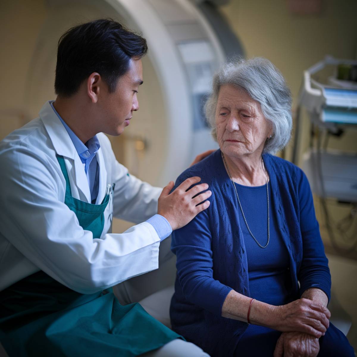
Patient Experiences With Angiography
In considering the patient-centered advantages of alternative techniques, it’s important to recognize the real-world experiences of those who undergo angiography. Patient testimonials often reveal insights that help us appreciate the nuances of this procedure. Here are some key themes from their feedback:
- Comfort levels: Most report minimal discomfort, largely due to local anesthesia at the catheter insertion site.
- Supportive staff: Many highlight how dedicated healthcare teams alleviate anxiety, reinforcing the importance of clear communication.
- Satisfaction: With 98.5% expressing favorable views, it’s clear that attendees value their experiences during the procedure.
Encounters with angiography often show that outcomes hinge not only on technical aspects but also on the emotional journeys patients undertake. By sharing their procedure feedback, these individuals illuminate how the blend of medical care and compassion leads to a more positive experience overall.
Future of Angiography

As we look to the future of angiography, we’ll witness remarkable advances in imaging technologies that promise to enhance patient safety considerably. Innovations like photon-counting CT and MRI angiography are setting a new standard, allowing for thorough assessments while minimizing risks associated with traditional methods. By embracing these advancements, we’re not only improving diagnostic accuracy but also ensuring a much safer experience for patients.
Advanced Imaging Technologies
While the landscape of angiography evolves, advanced imaging technologies are paving the way for groundbreaking diagnostic capabilities in the medical field. These innovations not only enhance image clarity but also contribute to significant diagnostic innovation. We’re witnessing an era where various modalities provide extensive insights into vascular health.
- CT Angiography: Visualizes blood vessels non-invasively using contrast dye.
- MRI Angiography: Assesses blood vessels without exposing patients to X-rays.
- Magnetic Particle Imaging (MPI): employs magnetic nanoparticles for tissue imaging.
Improved Patient Safety
Embracing innovative approaches to angiography enhances patient safety substantially, making it a cornerstone of modern medical practices. By implementing advanced CT scans, we substantially reduce breathing time, which not only minimizes exposure to complications but also enhances patient care. The improved visualization these techniques provide enables accurate blockage detection, lowering the risk of kidney injury and major heart complications. Additionally, combined procedures promote higher patient satisfaction and streamline healthcare delivery, optimizing safety protocols throughout our clinics. As we pioneer these advancements, we’re not only reducing the need for invasive procedures but also contributing to better long-term patient outcomes. Together, we’re building a safer, more effective angiography landscape for everyone involved.
Advances in Technology
Advancements in angiography technology are transforming the landscape of vascular imaging, enabling us to achieve remarkable image quality and diagnostic precision. As we explore the technological benefits, we see how these innovations enhance our ability to diagnose and treat vascular conditions effectively. Key progresses include:
- High-resolution imaging with CCTA, providing sub-millimeter spatial resolution for accurate coronary assessments.
- Non-invasive techniques, like CCTA, minimizing the need for traditional catheter-based procedures while still maintaining imaging efficacy.
- Integration of AI and machine learning to improve diagnostic capabilities and risk stratification.
These advancements not only refine the accuracy of angiography applications but also greatly reduce patient exposure to radiation, making the procedures safer and more effective. With tools like ECG gating and the Perivascular Fat Attenuation Index, we’re better equipped to evaluate and manage cardiovascular health. The future of angiography looks promising, marrying technology with patient care.
Angiography in Research and Development

As we explore the role of angiography in research and development, we can clearly see how advancements in imaging techniques enhance our ability to diagnose and treat vascular conditions. These innovations not only facilitate detailed insights into blood flow dynamics but also pave the way for more effective clinical trials that assess new therapies and improve patient outcomes. By examining these developments, we gain a deeper understanding of the potential angiography holds in shaping future medical practices.
Advancements in Imaging Techniques
While we’ve seen remarkable progress in imaging techniques, angiography continues to evolve, considerably enhancing our diagnostic and interventional capabilities. The landscape of angiography is shaped by imaging innovation and technology integration, which allow us to visualize vascular structures with unparalleled detail.
Key advancements include:
- Digital Subtraction Angiography (DSA): Clearer vessel visualization through background subtraction.
- Rotational Angiography: 3D imaging for improved decision-making in procedures.
- Dual-Energy CT: Enhanced tissue differentiation using photon-counting detectors.
These technologies not only improve patient outcomes but also streamline procedures, empowering us to make informed decisions swiftly. As we continue to innovate, the future of angiography looks promising, with endless possibilities for enhanced vascular health management.
Role in Clinical Trials
By integrating angiography into clinical trials, we greatly enhance our understanding of various vascular conditions and treatment methodologies. Angiography applications, particularly in stroke studies, allow us to visualize blockages and assess the efficacy of direct transfer protocols, such as the Direct to Angiography Suite (DTAS) trials, ultimately improving patient outcomes. In coronary artery disease research, Coronary Computed Tomography Angiography (CCTA) proves invaluable, offering high diagnostic accuracy and guiding treatment decisions. Additionally, interventional cardiology trials leverage early coronary angiography to safely identify acute occlusions. As we explore pulmonary and renal angiography studies, the insights gained pave the way for clinical trial innovations that refine our approach to managing complex vascular disorders, leading to tailored and effective patient care.
Frequently Asked Questions
What Should I Wear During an Angiography Procedure?
When preparing for an angiography procedure, we should wear loose, comfortable clothing that allows easy access to the area being examined. It’s important to avoid clothing with metal, as it may interfere with the imaging. We’ll also need to change into a hospital gown provided by the facility. Following these preparation tips helps guarantee a smooth process and keeps everyone focused on the procedure itself, making it a little less stressful.
Can I Eat or Drink Before Angiography?
Before our angiography procedure, we need to follow specific fasting guidelines. Typically, we should stop eating solid foods six hours prior, guaranteeing our bodies are clear for the test. Clear liquids might be acceptable up to two hours before. It’s crucial to adhere to our pre-procedure instructions, as this helps guarantee our safety and the procedure’s success. Let’s keep our healthcare provider informed about any allergies or concerns to minimize potential risks.
How Long Will the Recovery Take After Angiography?
After angiography, our recovery expectations usually range from one to two days for most of us to feel back to normal. However, we’ll need to monitor for any angiography complications, such as swelling or bruising, that may linger for a week or more. It’s crucial to keep the wound area dry and avoid strenuous activities to facilitate a smooth recovery. Regular follow-up checks guarantee we’re on the right track to complete healing.
Will I Feel Pain During the Angiography Procedure?
We recognize that you’re concerned about pain during the procedure. Most of us experience minimal discomfort, thanks to effective local anesthesia ensuring patient comfort. Initially, we might feel a brief stinging when the anesthetic is administered, followed by some pressure during catheter insertion. Post-procedure, we can manage any soreness with pain management strategies, like paracetamol. Remember, our priority is to make the experience as comfortable and worry-free as possible.
Can Angiography Results Be Seen Immediately?
Yes, we can often see angiography results immediately after the procedure. During types like coronary or peripheral angiography, the special X-ray machines provide real-time images. While this offers quick insights into blood vessel conditions, we must remember the angiography risks, such as allergic reactions to contrast dye or radiation exposure. Ultimately, immediate results help us assess blockages or abnormalities swiftly, leading to timely medical decisions that benefit our health.
Conclusion
To summarize, angiography serves as an essential tool in diagnosing and treating vascular conditions, connecting us to advancements in medical technology that enhance patient care. As we explore the future, we can’t overlook its role in innovative research and the continuous evolution of imaging techniques. Together, we’ll embrace the transformative power of angiography, ensuring that both we and our loved ones receive the best possible insights into our cardiovascular health.
References
- https://www.nhs.uk/conditions/angiography/
- https://top.ucsf.edu/procedure/angiography
- https://www.hopkinsmedicine.org/health/treatment-tests-and-therapies/angiogram
- https://www.cedars-sinai.org/programs/imaging-center/exams/interventional-radiology/angiography.html
- https://www.mayoclinic.org/tests-procedures/coronary-angiogram/about/pac-20384904
- https://en.wikipedia.org/wiki/Angiography
- https://www.medstarhealth.org/services/angiogram
- https://www.johnmuirhealth.com/services/cardiovascular-services/intervention/angiography-angiogram-arteriogram.html
- https://www.bmc.org/content/angiography
- https://medisimaging.com/academy/scientific-blogs/history-of-coronary-angiography-and-the-emergence-of-qfr/